There is no sufficient evidence to claim that air purifiers can help prevent transmission indoors. In fact, air purifiers may spread the virus faster. Why not just increase the ventilation by opening windows? This is a cheaper option rather than buying air purifiers.
“When a worker coughs or releases droplets near a colleague’s respiratory system, the droplets may spread throughout the call center via air flow from air purifier. In this fashion, a single infected person can give rise to an infection cluster. Attempts to prevent infection must not lead to new infections, and the installation of air purifiers may cause new problems. Therefore, using air purifiers to control the spread of COVID-19 should be approached with caution.”
(This is a review / perspective journal article. So far, I can’t find any sound evidence that tells me that air purifiers help prevent transmission.)
Personal thoughts on “COVID-19 reinfection” and CDC’s claims that patients are not infectious and have recovered 10 days after their symptoms disappear even if they may test positive in RT-PCR:
It is well-established in many studies that even recovered patients can test positive in RT-PCR and yet are not infectious [1]. Recovered patients can test positive in RT-PCR because RT-PCR only tells you that fragments of the virus are present. This has led the CDC to publish guidelines on discharging patients 10 days after symptoms have resolved [1].
It is also well-established that T-Cell responses are robust [2], but antibodies from B-Cells do not last for a long time [3]. Still, we’re still confident that T-Cell responses can kill the virus and prevent reinfection.
Now, let’s imagine a certain Patient X. Patient X got infected and recovered. Patient X tests positive in RT-PCR via nasopharyngeal swab, but we know that these are just the fragments of the virus. We know that the patient is not infectious.
Now, let’s say this Patient is not able to maintain his B-Cell and T-Cell response for a sufficiently long time. Cytotoxic T-cells did not kill the rogue cells infected by the virus. Antibodies from B-Cells declined as time passes by. Patient still follows social distancing, hand hygiene, use of face masks and shields.
A virus is not a living thing, so you can’t kill it. In Chinese, the word “virus” is translated as “sickness toxin” (病毒). Normal poisonous chemicals don’t replicate, but viruses do.
So if we understand virus as a “toxin” – dual nature of a virus as a “toxin” and a “pathogen”, then naturally, even residual doses of it can cause sickness. By residual doses, we’re not talking about the non-infectious fragments of a virus, but the virus as a whole, or its virulence / pathogenic factors. This can explain why many viral diseases like Measles, Ebola, Varicella, HIV, HPV, HSV have post-infectious latent periods and post-infectious complications (meaning, even if they aren’t “infected”, they still manifest the disease or its sequelae).
If getting “infected” means it’s from a “living pathogen”, and “recovered” means recovering because the “pathogen is dead”, then a person cannot be “infected” with nor “recovered” from a virus. Viruses are in a limbo; they are neither “alive” nor “dead” because they are not composed of cells; they are not “living”. Like what my classmate said, they are “necromancers or dark wizards of life itself” [4]. They cause mutations in the DNA that cause cancer and drive evolution.
Given this, it is very well possible that people get reinfected – just not in the way one might understand. This is consistent with sporadic news reports. We might have to think of “reinfection” as the inability to expel an “alive”, but non-living, “sickness toxin” from the body.
(Note that I currently don’t have any hard evidence to support my claims.)
The book “The Death of Cancer” by Dr. Vince DeVita really struck my prevailing pessimism towards late-stage cancer and further solidified my interest in cancer. As I read the book, I felt that there were too many good quotes that just had to be quoted here. Dr. DeVita is one of the authors of the world’s most credible textbook on Oncology and is internationally renowned for his contribution on the treatment of Hodgkin’s lymphoma.
Below is the interview wherein Dr. DeVita discusses his book.
Without further ado, here are the quotable quotes:
“Many people will tell you that treating patients with advanced cancer is not worth it. That’s baloney. When a doctor says that, what he usually means is that it would not be worth it for him. Doctors often have trouble walking in the shoes of their patients. If a patient is functional and there is a reasonable chance of a useful response, I believe that it is worth trying one or two cycles of treatment. That can take a month to a month and a half.” – Vince DeVita, MD (The Death of Cancer)
“I could have told you a story with a happy ending. I instead chose to tell you one that could have had a happy ending because it illustrates what has been, for me, a source of perennial frustration: at this date, we are not limited by the science; we are limited by our ability to make good use of the information and treatments we already have. Too often, lives are tragically ended not by cancer but by the bureaucracy that came with the nation’s investment in the war on cancer, by review boards, by the FDA, and by doctors who won’t stand by their patients or who are afraid to take a chance.” – Vince DeVita, MD (The Death of Cancer)
“As early as 1907, Paul Erhlich, the great German chemist, had begun to use rabbits with syphilis to test the efficacy of certain chemicals to fight infectious diseases. He thought about doing the same for cancer but was so pessimistic about the likelihood of success that he hung a sign over the door leading to the cancer lab. It read, “Give up all hope oh ye who enter.” Other doctors had tried a solution of arsenic, called Fowler’s solution, to treat leukemias and lymphomas, with no success.” – Vince DeVita, MD (The Death of Cancer)
“Halsted’s procedure lived up to its name. It was radical. It left a woman with only a thin skin graft covering the chest wall, through which her rib cage was clearly visible. Removing this much tissue also tended to cut off the drainage path for lymph channels—small, almost invisible vessels that drain excess fluid from organs, strain it through lymph nodes, and then deliver it back to the bloodstream. This blockage resulted in the additional disfigurement of a swollen arm.
Because Halsted was a surgeon of such renown, removing tumors en bloc became the standard for all cancer surgery. In fact, surgeons referred to it as “the cancer operation.
Applied to head and neck cancers by the famous head and neck surgeon Hayes Martin, it became the commando procedure, in which surgeons took the tumor, the lymph nodes, the strap muscles of the neck and the jawbone (which make it possible to speak, chew, and swallow), and even the tongue, if necessary. It was the most disfiguring operation ever invented. Patients wore special veils across their faces to hide their disfigurement.
Applied to colon and rectal cancers, it meant permanent colostomy. Applied to tumors on the extremities, such as bone cancers affecting the leg, it meant radical amputations, sometimes removing the entire femur bone from the hip on the affected side or taking off half the pelvis, too. Applied to pancreatic cancer, it became the Whipple procedure, which involved the removal of the pancreas, stomach, duodenum, spleen, and surrounding lymph glands.
Applied to cervical cancer, it became the Wertheim procedure, in which the surgeon totally excavated a woman’s pelvis, resulting in not only a colostomy but also the redirection of urine flow.” – Vince DeVita, MD (The Death of Cancer)
“Gilman and Goodman gave nitrogen mustard to rabbits and noted that the cells within the rabbits’ bone marrow and lymph nodes disappeared. It occurred to them that this might be a positive when it came to certain types of cancers, such as lymphomas, which inhabit the lymph nodes. When they tried nitrogen mustard on mice in which tumors had been transplanted, the cancer went away. The idea of giving nitrogen mustard to an actual person was a tantalizing next step.
The doctors couldn’t specify the drug, nitrogen mustard—similar to mustard gas—because it was being studied in secret as part of the war effort. But that’s what they were referring to.” – Vince DeVita, MD (The Death of Cancer)
“The ultimate outcome, in all cases, was the same. The children died. Leukemia was a death sentence, a torment to the children who had it, their parents, “and the doctors who had to stand by, knowing full well they were doing things that would not work, while the children slipped away.
It was in this atmosphere that Frei and Freireich announced that they planned to use chemotherapy to cure childhood leukemia, which elicited ridicule because everyone knew it was impossible. And then they’d come up with an even more radical idea than giving toxic drugs to children who were, quite literally, on their deathbeds. They decided to give the drugs in combination—two or more at a time. The medical community was scandalized. Using more than one drug at a time to treat something was, as a general rule, considered sloppy medicine. Using more than one highly toxic drug, in children, no less, and in children who were already suffering, was unfathomable.” – Vince DeVita, MD (The Death of Cancer)
“I wasn’t sure if these scientists were maniacs or geniuses. But most of the senior doctors at other NIH institutes had long since decided on the former. “Every Wednesday, in the NCI solarium, there were grand rounds for the entire hospital, where scientists would stand at the podium and discuss their work while their peers listened and, perhaps, gave helpful feedback. Whenever Frei and Freireich were featured, it was a verbal bloodbath.
Some of the doctors in the audience would scream out interjections while Frei and Freireich talked. “This is a meat market! What a butcher shop!” I saw it happen many times. Frei and Freireich ignored them, but it was embarrassing and shocking to watch. Never had I seen doctors behave this way toward other doctors. And it made us clinical associates question what we were doing. Were we doing the right thing? Were we accessories to what other doctors were portraying as unethical behavior? How could we know? And what was our recourse?” – Vince DeVita, MD (The Death of Cancer)
“One of the main obstacles to treating leukemia patients with chemotherapy, for instance, was that they often succumbed to infection before we could even try to treat them with drugs. They were uniquely vulnerable to infection because cancer cells crowded out their infection-fighting white cells and lymphocytes, which control the immune system. One infection they commonly fell prey to was pseudomonas meningitis, which was quickly fatal once it set in. There was no IV antibiotic known to be able to cross the blood-brain barrier, a blockade made mostly of complex lipids that few drugs seemed able to penetrate, in order to get to the bacteria.
Freireich instructed us to get around that problem by injecting an antibiotic called polymyxin B directly into the spinal fluid intrathecally—that is, via a lumbar puncture in the lower back. This was something the label on the drug vial expressly instructed you not to do. The first time Freireich told me to do it, I held up the vial and showed him the label, thinking that he’d possibly missed something. “It says right on there, ‘Do not use intrathecally,’” I said. Freireich glowered at me and pointed a long bony finger in my face. “Do it!” he barked. I did it, though I was terrified. But it worked every time.
When pseudomonas causes an infection in a normal patient, it creates an abscess, a pus-filled mass made up of millions of white blood cells. Leukemic patients didn’t get abscesses, Freireich said, because they didn’t have enough normal white cells to form one. If you saw a kid with a fever, you couldn’t rule out pseudomonas aeruginosa in less than a couple of days. And once pseudomonas was in the bloodstream, it was usually fatal in leukemic kids in twenty-four to forty-eight hours. Freireich wanted us to treat it as soon as we suspected it was what we might be looking at. And, because we couldn’t be sure what bacteria was responsible, he insisted we use several antibiotics at a time, to cover all possible bases.
In medical school and during our early training, we’d been taught to culture the blood for bacteria, find the exact bacterial culprit, and, only then, treat the specific cause of the infection with the right antibiotic. Treating with more than one drug, without identifying a specific cause, was not standard practice. But Freireich insisted on it. The problem, Freireich said, was that as in the case of pseudomonas aeruginosa, it took days for bacteria cultures to grow, and we didn’t have the luxury of time with our leukemic patients, who had few or no white blood cells and could die within hours from an untreated infection. “We can always back away later,” he said.” – Vince DeVita, MD (The Death of Cancer)
“Until the turn of the twentieth century, the assumption was that Hodgkin’s was a form of tuberculosis. It wasn’t an unreasonable assumption. One subtle aspect of Hodgkin’s is that even patients with early-stage disease are susceptible to infections, particularly tuberculosis and fungal infections. Ergo many patients with Hodgkin’s also had tuberculosis, which was nearly epidemic in the early nineteenth century. One well-known pathology textbook dating to 1919 said, “Tuberculosis follows Hodgkin’s disease like a shadow.”
It wasn’t until 1902 that Drs. Dorothy Reed and Carl Sternberg at Johns Hopkins University, aided by microscopes, were able to look at cells that came from the lymph nodes of people with Hodgkin’s. Most cells, even cancer cells, have just one nucleus. Reed and Sternberg saw something unusual—a cell with two, looking back at them like the eyes of an owl.
It was Reed and Sternberg who reclassified Hodgkin’s as cancer. The owlish-looking cell came to be known as the Reed-Sternberg cell. (In medicine, first spotters get naming rights. The disease was named after Hodgkin, but Reed and Sternberg had been the first to spot the cell.) Not all malignant cells in Hodgkin’s look like Reed-Sternberg cells. But in order for a lymphoma to be classified as Hodgkin’s, they have to be there.” – Vince DeVita, MD (The Death of Cancer)
“Medical school doesn’t prepare you to speak with patients about their impending deaths. And even though I was now compelled to deal with death quite a lot, I hadn’t gotten past my discomfort.” – Vince DeVita, MD (The Death of Cancer)
“I’d learned that in order to do anything outside the mores of medicine, you needed someone in a high place, or at least high enough, to run interference.” – Vince DeVita, MD (The Death of Cancer)
“A new pair of eyes often sees something obvious that the primary doctor, staring at the chart and the patient every day, has missed.” – Vince DeVita, MD (The Death of Cancer)
“Radiotherapists began to lose referrals as the management of the disease shifted to medical oncologists. As a result, many began to refuse to cooperate in large studies of early-stage patients unless radiotherapy was included in every group of each study, which meant that you could no longer gauge the outcomes with and without radiotherapy.
One colleague, the leader of the largest Hodgkin’s disease clinical trials group in the world, told me privately that he had to include radiotherapy in each group of patients with early disease if he wanted to accrue patients. He needed the cooperation of radiotherapists who often saw these patients first. Ergo, many patients were receiving radiotherapy—needlessly.
This assured that no matter the outcome, radiotherapists would always have a role in the care of patients with Hodgkin’s disease—even though our data already indicated that it wasn’t necessary and there was reason to believe that exposure to radiation could put patients at risk of future cancers down the road.” – Vince DeVita, MD (The Death of Cancer)
“A good cancer doctor never wants to be too far from his pathologist.” – Vince DeVita, MD (The Death of Cancer)
“In 1946, the microbiologist Selman Waksman published a paper on streptomycin, a new class of antibiotic that appeared to be effective against certain microbes that were not vulnerable to penicillin. Mary, who was not a scientist, pointed out to Waksman that the drug had had some effect against tuberculosis. There were no useful drugs for the disease at the time, and Waksman himself, along with most of organized medicine, initially thought the results not promising enough to pursue. Mary’s instincts said otherwise. She persuaded Waksman and the Merck pharmaceutical company to support tests of streptomycin against TB. By 1952, the widespread use of streptomycin resulted in cutting mortality from TB in half—only a few years after the drug’s discovery. You might say she helped him win the Nobel Prize, which he received that same year for developing the tuberculosis cure.” – Vince DeVita, MD (The Death of Cancer)
“Her mantra was that Congress wouldn’t be generous in funding a concept—something like “biological research.” But propose funding for an institute named after a feared disease, and you’d make progress.” – Vince DeVita, MD (The Death of Cancer)
“What separates great doctors from mediocre doctors is the ability to integrate all the information they see in their patients, on the wards, on a day-to-day basis and understand it.” – Vince DeVita, MD (The Death of Cancer)
“All researchers can use another pair of eyes—or a dozen pairs—to make sure they stay on the right track.” – Vince DeVita, MD (The Death of Cancer)
“Not all stories had happy endings, but it was still a privilege to be a part of them. […] But they meant something, these moments. They not only taught you what it means to be a doctor, and a human being, but gave you opportunities to step up and be one.” – Vince DeVita, MD (The Death of Cancer)
“Jim Watson, of double helix fame, had been appointed to the NCAB because he was a brilliant scientist. But he also hated the concept of the war on cancer and was purposefully rude at board meetings to show his contempt. The board chair couldn’t control him. Watson was, after all, a Nobel laureate. He liked to lean back and put his feet up on the table and read The New York Times, lowering the paper only to interject a derogatory comment here and there. Schmidt patiently waited for the board chair to say something, but he didn’t. On one occasion, when the board was discussing the newly formed Cancer Centers Program, Watson lowered the paper and said, “This is a pile of shit.”
Benno asked for the floor and said, “I have the budget of the centers program here.” He then noted that one of the largest grants went to “Dr. Watson at Cold Spring Harbor.” “Are you part of the pile of shit, Dr. Watson?” A stunned Jim Watson put the paper and his feet down and remained silent. His bluff had been called. Then Schmidt got in his limo and went to the White House and asked that a disruptive Jim Watson be removed from the board. Watson’s six-year appointment was terminated after two years. You didn’t want to mess with Benno Schmidt, even if you had a Nobel Prize. In 2009, Jim wrote an editorial in The New York Times claiming that he had been booted because “the NCI went clinical,” which wasn’t even close to being true. Then and now, about 85 percent of the NCI’s budget went to supporting basic research.” – Vince DeVita, MD (The Death of Cancer)
“Bernie’s study was controversial, because breast surgeons made their living doing radical or total mastectomies, and they did not want to hear that that was no longer necessary. […] Not because the science was bad, but because they didn’t want to give up the more extensive surgery that created 90 percent of their income. It was economics. […] Most surgeons and radiotherapists would never admit that they opposed Fisher’s findings because they threatened the doctors’ incomes. Instead, they questioned his integrity.” – Vince DeVita, MD (The Death of Cancer)
“It is unrealistic to use survival as an end point. Consider the number of cancer cells a drug has to kill to be effective. The average patient with advanced cancer has more than a kilogram of tumor on board. That’s many billions of cancer cells, because a lump about one centimeter across contains roughly one billion cells. Even after surgery, when a primary cancer has been removed and no errant cells can be detected, a patient can have more than a billion cancer cells circulating around the body.
To see a complete response to treatment, a drug has to kill 99.99 percent of the cancer cells. And then a very curious thing happens.
The time it takes for the surviving tumor cells to grow back to their pretreatment levels varies depending on the number of cells that have survived the treatment. Smaller populations grow back faster than larger populations. It seems counterintuitive, but it’s true. The net effect is that if you have two treatments—one that reduces the cancer cell population in a patient to just a few cells, and one that reduces the population to one billion cells—neither patient will have visible tumor masses at that point. But after five years, both will have the same number of cancer cells present, as the rapidly growing smaller population catches up. The five-year survival rates will be the same. But the treatment that reduced the population to a few cells is the one you want to go forward with. But which one is that? You cannot tell by using five-year survival rates as an end point.
This is called the Norton-Simon effect, named after two of my colleagues who discovered it. This alone should lead to the reconsideration of the use of survival rates in advanced cancer patients as a requirement for approval of a new drug.” – Vince DeVita, MD (The Death of Cancer)
“Cancer is the most curable chronic disease. But it is more often fatal as well.” – Vince DeVita, MD (The Death of Cancer)
“So when I say we want to convert most cancers into chronic diseases, I mean we want cancer patients we can’t cure to more closely resemble those with diabetes or arthritis—to live out a normal or near-normal life span while managing the condition. Immortality is not our goal here, just a normal life, free from the gnawing worry that a premature death is lurking just around the corner.” – Vince DeVita, MD (The Death of Cancer)
“As a result of the war on cancer, the cancer cell is no longer a black box; it’s a blueprint. And we can read blueprints. We understand the cancer cell’s stages, how it thinks, what drives it. And we have the tools to attack each of the steps on the way to malignancy. There isn’t a question about cancer we can’t address, at this point, without the expectation of a usable answer.” – Vince DeVita, MD (The Death of Cancer)
“As optimistic as I am, I don’t think there will ever be a world in which cancer doesn’t occur. It’s in our biology. In the millions of cell divisions that take place in our bodies every day, there are too many opportunities for mistakes. And some of those mistakes will activate a cancer hallmark and give rise to cancer.
We can prevent it, to some extent, by avoiding the things that make those mistakes more prone to happen, like smoking. And we can take advantage of medication that seems, in some cases, to prevent those mistakes.” – Vince DeVita, MD (The Death of Cancer)
“It is now clear that the cancer cell is recapitulating developmental biology. In plainer terms, it thinks it’s a fertilized egg on its way to becoming an embryo, and it uses the powerful growth machinery of the early embryo, normally shut down in adulthood, to fire its growth and development. You can look at cancer, then, as a failure in the control of the delicate cellular machinery that evolved to prevent embryos from becoming monsters. This sounds fantastic, I know, but it’s true. There are cancers that, when removed, are found to have hair and teeth and other tissues. They’re called teratomas, from the Greek word teras—meaning monster.” – Vince DeVita, MD (The Death of Cancer)
“The real toxicity of low-dose chemotherapy is premature death. If your doctor recommends it, head for the nearest exit.” – Vince DeVita, MD (The Death of Cancer)
“Frank Schabel, who worked at the Southern Research Institute in Birmingham, Alabama, with Howard Skipper, did a set of experiments that were very telling about the effect of full doses of chemotherapy on the ability to cure a metastatic tumor. He implanted mouse sarcomas into groups of mice and watched the sarcomas grow until the mice had lots of measurable tumors in their lungs. Then he treated them with combinations of cancer drugs. At full doses, all of the mice went into complete remission. That is, all of their measurable tumors disappeared. Then he stopped treatment and followed the mice. Within months, about half of the mice developed recurrences and died. The remainder were cured.
So he had a 100 percent complete remission rate and a 50 percent cure rate. Then he began to reduce the doses of the drugs. He found that if he reduced the doses of the drugs by 20 percent, he still got a 100 percent complete remission rate. But he got only a 25 percent cure rate.” – Vince DeVita, MD (The Death of Cancer)
“Dr. Stanley Cohen at Vanderbilt University was a basic scientist interested in cell growth. In the course of his experiments, he had injected newborn mice with extracts from salivary glands. He was surprised when the newborn mice prematurely opened their eyes and erupted teeth. Something in the salivary extracts was speeding cell growth.
Ultimately, Cohen figured out that the material he was purifying was accelerating the growth and separation of epithelial cells that sealed the mouse eyelids and caused teeth to sprout. He discovered, isolated, purified, and sequenced a material he called epidermal growth factor (EGF), a protein that stimulates the growth of epithelial and other cells and enhances certain developmental growth.
He was also able to identify the target receptor for EGF and the mechanism of its action, providing a breakthrough in understanding how signals from outside a cell reach the inside of a cell, how cells talk to one another.
This turned out to be one of the most important discoveries of the twentieth century. It has had an important impact not only on the understanding of how cancers grow but also on cancer treatment. The discovery enabled scientists to further explore the cell growth process set in motion by EGF stimulation and to develop drugs to block the individual steps. The EGF receptor is expressed in virtually every cell in the body, and it is often over-expressed in cancer cells. This realization led to a huge increase in research on targeted therapies. In 1986, Cohen and Levi-Montalcini shared the Nobel Prize for identifying the first growth factors.
The biotech firm ImClone was one of the companies to take up the challenge of finding drugs to block EGF. I’m quite familiar with this work, because I was on the company’s board when this research was being done. Researchers identified four EGF receptors—labeled EGF-1 through EGF-4. ImClone developed an antibody, called Erbitux, to block the EGF-1 receptor, the most common variety. The drug proved to be useful in prolonging survival in both colon and lung cancer patients. And its side effects on normal cells were trivial: a rash around hair follicles. (Oddly enough, people who got a rash responded to the drug more often than those who didn’t.) Others discovered that EGF-2, also known as HER2, is over-expressed in about a third of patients with breast cancer. And it is not commonly expressed in normal cells. Another drug, Herceptin, which blocks the HER2 receptor, is a mainstay of the treatment of breast cancer.” – Vince DeVita, MD (The Death of Cancer)
“The death of cancer is inevitable. It is a question not of if but of when.” – Vince DeVita, MD (The Death of Cancer)
“The rate-limiting step in eradicating cancer today is not the science but the regulatory environment we work in.” – Vince DeVita, MD (The Death of Cancer)
Stroke occurs when a blood vessel that carries oxygen and nutrients to the brain is either blocked by a clot or bursts (or ruptures) [American Stroke Association].
If a blood vessel gets blocked by a clot, it’s called “Ischemic Stroke”.
If a blood vessel ruptures, it’s called a “Hemorrhagic Stroke” or “Intracerebral Bleed” (blood inside the brain).
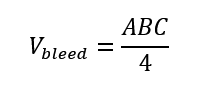
Traditionally, if stroke occurs, patients undergo a CT Scan of the head. Afterwards, neurologists (doctors who specialize on diseases of the nervous system) determine if it is one type or the other. If it is a bleed, then they calculate the volume of the bleed using a formula called “Kothari Formula”. Why is this important? The prognosis (chance of a patient living or dying) can be predicted from the volume of the bleed!
During my Neurology rotation, we used a guidebook titled “Ictus” which contains much of the pertinent information used to manage the most common neurological diseases seen in the hospital. Stroke is the most common one among them. The guidebook elucidated that the volume of an intracerebral bleed using the Kothari Formula is as follows:
When I saw this formula, I could not accept such a simplistic ‘estimate’ formula, especially with the denominator of 4. The volume of a bleed has spherical elements, so I believe that the Greek letter π should at least be part of the formula.
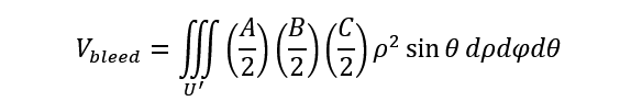
Consequently, I tried to derive the formula of the bleed using calculus.
Let there be an ellipsoid with dimensions A/2, B/2, C/2 satisfying the equation:
First, we have to use generalized spherical coordinates. Let:
Since the absolute value of the Jacobian for transformation of Cartesian coordinates into generalized spherical coordinates is:
Hence,
The volume of the ellipsoid is expressed through the triple integral:
By symmetry, we can find the volume of the 1/8 part of the ellipsoid lying in the first octant (x ≥ 0, y ≥ 0, z ≥ 0) and then multiple the result by 8. The generalized spherical coordinates will range within the limits:
Then the volume of the ellipsoid is:
When I saw the result, I was even more confused. How can the calculated theoretical volume be almost twice as large as the one shown in the guidebook? Were we overestimating the volume of the bleed?
I started comparing it with other methods of getting volume of solids:
Volume of Prism = ABC
Volume of Elliptical Cylinder = πABC
Volume of Pyramid = ABC/3
Volume of Elliptical Cone = πABC/3
I started researching for journals online. Among the references I noted [1][2], none of them pointed to ABC/4. My derivation is closest to ABC/2, with π being approximately 3 – this is more consistent with the original research [1] made by Kothari himself. Another paper [2] found out that the true volume is closer to ABC/3 – the volume of a pyramid.
Again, none of the papers I’ve seen so far show ABC/4.
I cannot accept this formula yet.
Maybe, the discrepancies can be explained by the following points:
Perhaps, just as the papers indicated, the reason behind this formula lies within the radiologic and technical aspects of the CT Scan used – the resolution and the size of the slices.
When a bleed occurs, it occurs inside the brain. There are surrounding structures that change the shape of the bleed.
The hemodynamics and pressure dynamics inside the brain are defined by the Monro-Kellie Doctrine – The total pressure inside the cranium is fixed; an increase in one of the three components of the brain, namely, brain tissue, blood, and cerebrospinal fluid (CSF) would occur at the expense of another. A bleed can occur at first, but the edema (brain swelling) that occurs later can therefore further decrease the volume.
This begs the research question: “What if we construct a formula – a multivariate function – of the volume of intracerebral bleed as a function of time as well as blood pressure?”
References
[1] Kothari RU, Brott T, Broderick JP, Barsan WG, Sauerbeck LR, Zuccarello M, Khoury J. The ABCs of measuring intracerebral hemorrhage volumes. Stroke. 27 (8): 1304-5. Pubmed
[2] Huttner HB, Steiner T, Hartmann M, Köhrmann M, Juettler E, Mueller S, Wikner J, Meyding-Lamade U, Schramm P, Schwab S, Schellinger PD. Comparison of ABC/2 estimation technique to computer-assisted planimetric analysis in warfarin-related intracerebral parenchymal hemorrhage. Stroke. 37 (2): 404-8. doi:10.1161/01.STR.0000198806.67472.5c – Pubmed .
Life is short. Somebody can seem perfectly stable and then suddenly crash and die.
I hadn’t gotten time to process things because I had to immediately line (give fluids) another patient who is also dying in order to save his life.
I couldn’t help but be enraged at the morally decadent people on top who live without worries, leaving PGH in such a depraved state with no water, lack of adequately-sized IV cannulas, lack of tubes to contain blood, overcrowded emergency room owing to an inefficient referral system, and overworked nurses and doctors. Health care workers rely on their own stash of materials which are barely enough.
And this isn’t the complete picture. The remaining 70% of Filipinos who die without medical attention are definitely not in any hospital (not just PGH). While hospitals in Japan, US, Europe, and the rest of the world are focused on developing the latest technology to cure HIV-AIDS and cancer, we couldn’t even maintain water and medical equipment supply in our own national hospital.
“Welcome to PGH,” the nurse told me.
(And I deeply respect this nurse because not only is she able to do IV lining on difficult patients but she also gifted me a roll of micropore tape. She also taught me how to troubleshoot a leaky foley catheter.)
(And there’s also another nurse whom I also respect deeply because despite the lack of properly-sized IV cannulas (pink and blue), he was able to insert extra-large gauge 16 IV cannulas (gray) with ease on patients, allowing a sufficient flow of IV fluids, saving their lives! Nurses actually save more lives than doctors!)
A part of me couldn’t help but blame myself because I felt that I didn’t give more attention to that patient when she suddenly said that her neck is very painful as she lost control of her neck right before she crashed and died. Even so, would I have been able to save her on time? Would I have had given her more attention, and perhaps saved her, if it weren’t for the tons of paperwork to accomplish and the demand to monitor a lot of patients? Am I not being too hard on myself?
Certainly, if this patient was seen, given proper attention, and operated on time, she would have a lot, lot more years to live.
That woman in her early thirties seemed stable (but is hypotensive), and she even talked while I extracted blood and inserted a nasogastric tube. Later, I had to do CPR – exerting force on my hands pushing downward towards the chest of the dying patient’s chest who was alive just moments ago.
“Just moments ago, she was alive.”
Such a grueling thought gnaws away my rational and calm self.
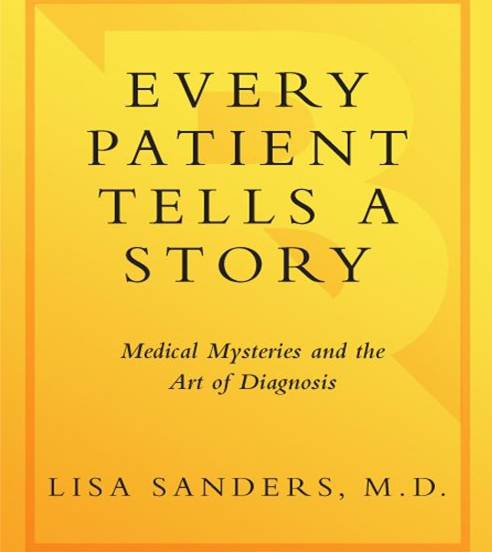
Every Patient Tells A Story is a book written by Dr. Lisa Sanders, one of the medical consultants of the popular television series House MD.
In this book, she talks of interesting cases such as Wilson Disease, Rocky Mountain Spotted Fever, Lemierre Disease, Lyme Disease, Thoracic Outlet Syndrome, Sjögren Syndrome, Still’s Disease, Giant Cell Arteritis, and IPEX Syndrome (Immunodeficiency, Polyendocrinopathy, X-Linked). She even mentioned a case wherein a chief complaint of forgetfulness was due to a paraneoplastic syndrome (due to hormone/s produced by malignant cancer cells).
Consequently, she echoed the paramount importance of history taking and physical examination, asserting that not even the CT Scan could fully replace the Stethoscope.
She also discussed the fact that neither Google nor any other diagnostic software could accurately diagnose the more common diseases. However, Google, being free and accessible is actually the best diagnostic tool for rarer diseases with unusual manifestations. She believes that no software can ever replace the medical doctor.
Here are some notable quotes from the book:
“I’d like to mention—endocarditis, tuberculosis, Wegener’s granulomatosis, Kawasaki’s aortitis, Jakob-Creutzfeldt dementia, and eosinophilic gastritis.” She rushed through this list of arcane diseases and ended with a laugh. “I don’t know any of the cases I’m about to hear but there’s a darn good chance I’ve mentioned at least one case diagnosis in that list.” – Dr. Faith Fitzergald (Every Patient Tells a Story)
“In this forum, even if you don’t ultimately figure out the case, you get credit for having the final diagnosis among the diseases you considered on the way. Fitzgerald was acknowledging that the cases she would be likely to confront that day would not be the same as those doctors routinely see in daily practice. Instead they would be the “fascinomas,” the intriguing cases physicians share at the watercooler, the nurses’ station, or in hospital stairwells.” – Dr. Lisa Sanders (Every Patient Tells a Story)
“Doctors build a story about the patient in order to make a diagnosis. It is a story based on the patient’s story but it is freed of most of the particular details of the individual, and structured to allow the recognizable pattern of the illness to be seen.” – Dr. Lisa Sanders (Every Patient Tells a Story)
“You’re starting out on the journey across this bridge, this education, and right now you are on the same side as your patients. And as you get halfway over the bridge you’ll find yourself changing and the language the patient had and you had is being replaced by this other language, the language of medicine. Their personal story is being replaced by the medical story. And then you find yourself on the other side of that bridge—you’re part of the medical culture. When you get there, I want you to hold on to every bit of your old self, your now self. I want you to remember these patients.” – Dr. Lisa Sanders (Every Patient Tells a Story)
“Medicine—to the extent that it can be called a science—is a sensual science, one in which we collect data about a patient through touch and the other senses according to a systematic method in order to make a diagnosis. Most patients are willing to be touched by their doctor. They expect it.” – Dr. Lisa Sanders (Every Patient Tells a Story)
“In medical school, starting with anatomy class, doctors are taught to understand the body by taking it apart, one piece at a time. What you walk away with, at the minimum, is an uncanny ability to objectify the hell out of even the most intimate body parts. For anyone else, this might be considered disrespectful, but for doctors, a clinical and objective view of, say, a female breast offers us the chance to see it isolated from its other, often sexual, contexts. We are taught to handle a breast as a separate object.” – Dr. Lisa Sanders (Every Patient Tells a Story)
“The sicker the patient, the greater the temptation to skip the fundamentals—like the physical examination—and to rely on the available technology to provide us with answers. It’s a temptation that can sometimes prove fatal” – Dr. Lisa Sanders (Every Patient Tells a Story)
“Those minutes of terror and confusion I felt standing powerless in her room served as a visceral reminder throughout my training (and even now, occasionally) that the big picture isn’t enough in medicine; that the overall impression of a patient is worthless without looking further and paying attention to the specific measurements of health or sickness that were behind the impression in the first place.” – Dr. Lisa Sanders (Every Patient Tells a Story)
“You mustn’t avert your eyes from abnormality. You need to seek it out. You need to figure it out. And it doesn’t just turn off when you leave your office.” – Dr. Lisa Sanders (Every Patient Tells a Story)
“We have tremendous faith in our ability to see what is in front of our eyes. And yet the world provides us with millions of examples that this is not the case. How often have you been unsuccessful in looking for an object and recruited the help of someone who finds it immediately right in front of you? Or had the embarrassing encounter with a friend who confronts you angrily after you “ignored” his wave the night before while scanning for an open seat in a crowded movie theater?” – Dr. Lisa Sanders (Every Patient Tells a Story)
“In many ways, the heart exam stands as a symbol of the entire physical exam. It’s not the most complicated exam—the neurological exam is the probably the most complex. Nor is it the most technically difficult exam—looking at the retina of the eye may get that honor. And it’s not the most time-consuming exam—that would probably be the psychiatric exam. But the heart exam was the first examination developed in modern medicine and the one most strongly linked with the physician’s role as diagnostician and caregiver.” – Dr. Lisa Sanders (Every Patient Tells a Story)
“Testing has changed how medicine is practiced. Doctors can now be far more certain of a given diagnosis with the help of tests than ever before in the long history of medicine.” – Dr. Lisa Sanders (Every Patient Tells a Story)
“Faulty knowledge was the key factor in only a few of the missed diagnoses, each of which involved a rare condition. Faulty data gathering—an inadequate history, missed findings on the physical exam, or misinterpreted test results—was a more common problem, playing a role in 14 percent of the diagnostic errors. Faulty synthesis—difficulty putting the collected data and knowledge all together—by comparison, played a role in well over half of the incorrect or delayed diagnoses.” – Dr. Lisa Sanders (Every Patient Tells a Story)
“Because Google is so universally available, simple, fast, and free, it may become the go-to diagnostic aid for oddball cases. Even the august New England Journal of Medicine finds Google “helpful in diagnosing difficult and rare cases.” Google gives users ready access to more than three billion articles on the Web and is far more frequently used than PubMed for retrieving medical articles.” – Dr. Lisa Sanders (Every Patient Tells a Story)
“Would a kind of super-efficient, integrated, intelligent computer system eliminate all diagnostic challenges? Would it replace doctors? Hardly. I believe the process of diagnosis will be made more effective and that it will be faster and easier in the future to zero in on what’s really wrong with a patient. But there will always be choices to make—between possible diagnoses, between tests to order, and between treatment options. Only a skilled and knowledgeable human can make those kinds of decisions.” – Dr. Lisa Sanders (Every Patient Tells a Story)
If I’d be asked which disease I think is more evil than cancer, I’d say it’s Idiopathic Pulmonary Fibrosis (IPF), and not cancer or any inborn disease.
Why? Why do I think it is worse than cancer or any inborn disease?
There’s no doubt that cancer is an evil disease. Breast, ovarian, lung, colorectal, and other forms of cancers have generally low survival rates if not treated. Breast cancer usually has the highest survival rate, while lung cancer has the lowest survival rate. However, the fact remains that these can be prevented by having a better lifestyle, not smoking, etc. In the recent era, many drugs have been developed to cure cancer. Usually, cancer is curable when discovered at an early stage, or even at Stage 4, as long as it is of the not-so-deadly type.
There’s also no doubt that inborn diseases like Down Syndrome, Pompe’s Disease, Edwards Syndrome, and others are very life threatening. Patients usually die early in life. However, most of these diseases are super rare, and babies born with these are not usually aware of the suffering they are undergoing.
Meanwhile, Idiopathic Pulmonary Fibrosis is a disease where your lungs over a long course of time become filled with bumps and scars. It has a higher chance of death compared to almost all kinds of cancer (except lung cancer). On top of that, doctors don’t know how the disease develops or works. That is why there is no cure for IPF. The patient just gets slowly and slowly suffocated from the inside. Unless lung transplantation is done, the patient will die. Unfortunately, finding lung donors is very, very difficult.
Still, if I were to be asked what disease is truly the most evil, my answer would be the Neglected Tropical Diseases (NTDs) such as Dengue and Schistosomiasis, as well as Tuberculosis. These are diseases of poverty and are a manifestation of poor public health measures. Deaths from these diseases are very preventable. Poor access to health services and lack of good governance indirectly kill people through these diseases.


